
Description
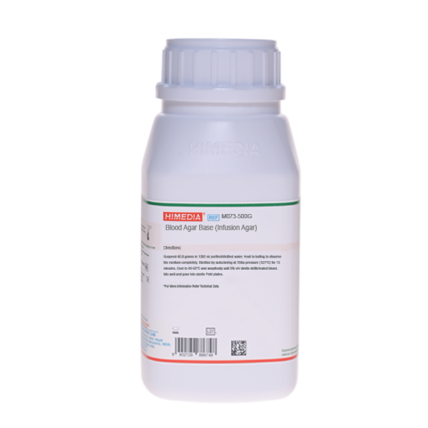
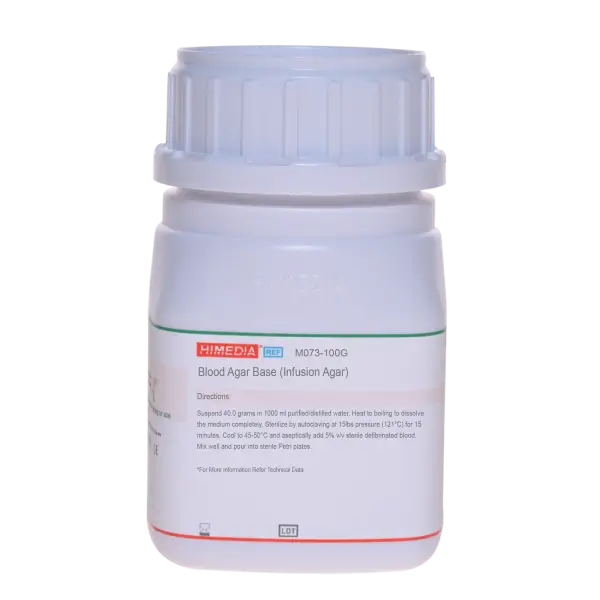
Blood Agar Base M073 Himedia
Blood Agar Base M073 Himedia adalah media pertumbuhan mikrobiologi standar berkualitas tinggi yang dirancang khusus sebagai substrat dasar untuk isolasi, kultivasi, dan penentuan sifat hemolitik dari berbagai jenis mikroorganisme patogen, seperti Streptococcus, Staphylococcus, dan bakteri rewel (fastidious) lainnya.
Dengan formulasi nutrisi yang kaya, media dehidrasi (powder) dari Himedia ini menjadi pilihan utama bagi laboratorium klinik, rumah sakit, lembaga penelitian, dan industri farmasi untuk memastikan hasil kultur yang akurat dan konsisten.
Spesifikasi Produk
| Fitur | Detail Spesifikasi |
| Nama Produk | Blood Agar Base |
| Manufaktur / Brand | HiMedia Laboratories |
| Kode Katalog | M073 |
| Bentuk | Bubuk Dehidrasi (Homogen, Krem hingga Kuning) |
| Kemasan | 500 Gram |
| Aplikasi | Isolasi dan kultivasi mikroorganisme fastidious serta uji hemolisis (setelah ditambah darah) |
Keunggulan Blood Agar Base M073 HiMedia
-
Nutrisi Maksimal untuk Bakteri Fastidious: Mengandung kombinasi pepton khusus dan ekstrak liver yang menyediakan nitrogen, vitamin, dan asam amino esensial untuk mendukung pertumbuhan bakteri yang sulit tumbuh.
-
Zona Hemolisis yang Jelas: Memiliki kejernihan basa yang sangat baik setelah ditambahkan darah steril (seperti darah domba atau kuda 5-10%), memudahkan identifikasi reaksi Alfa, Beta, atau Gamma hemolisis.
-
Kualitas Konsisten (Batch-to-Batch): Diproduksi dengan standar kontrol kualitas ISO yang ketat untuk memastikan hasil replikasi kultur yang andal di setiap pengujian.
-
Fleksibilitas Penggunaan: Selain sebagai media dasar Blood Agar, M073 juga dapat digunakan sebagai Nutrient Agar kaya nutrisi tanpa penambahan darah, atau diubah menjadi Chocolate Agar.
Composition
| Ingredients | g/L |
|---|---|
| HM peptone B# | 10.000 |
| Tryptose | 10.000 |
| Sodium chloride | 5.000 |
| Agar | 15.000 |
Quality Control
Appearance: Cream to yellow homogeneous free flowing powder
Gelling: Firm, comparable with 1.5% Agar gel
Colour and Clarity of prepared medium: Basal medium: Light amber coloured clear to slightly opalescent gel. After addition of 5% v/v sterile defibrinated blood : Cherry red coloured opaque gel forms in Petri plates.
Reaction: Reaction of 4.0% w/v aqueous solution at 25°C. pH: 7.3±0.2
pH: 7.10-7.50
Cultural Response: Cultural characteristics observed with added 5% v/v sterile defibrinated blood,after an incubation at 35-37°C for 18-48 hours.
| Organism | Inoculum (CFU) | Growth w/o blood | Recovery w/o blood | Growth with blood | Recovery with blood | Haemolysis |
|---|---|---|---|---|---|---|
| Neisseria meningitidis ATCC 13090 | 50-100 | fair | 40-50% | luxuriant | >=70% | none |
| Staphylococcus aureus subsp. aureus ATCC 25923 (00034*) | 50-100 | good | 50-70% | luxuriant | >=70% | beta |
| Staphylococcus epidermidis ATCC 12228 (00036*) | 50-100 | good | 50-70% | luxuriant | >=70% | none |
| Streptococcus pneumoniae ATCC 6303 | 50-100 | fair-good | 40-50% | luxuriant | >=70% | alpha |
| Streptococcus pyogenes ATCC 19615 | 50-100 | fair-good | 40-50% | luxuriant | >=70% | beta |
Key: (*) Corresponding WDCM numbers.
Storage and Shelf Life
Store between 10-30°C in a tightly closed container and the prepared medium at 2 – 8°C. Use before expiry date on the label.
On opening, product should be properly stored dry, after tightly capping the bottle in order to prevent lump formation due to the hygroscopic nature of the product. Improper storage of the product may lead to lump formation. Store in dry ventilated area protected from extremes of temperature and sources of ignition. Seal the container tightly after use. Product performance is best if used within stated expiry period.
Disposal
User must ensure safe disposal by autoclaving and/or incineration of used or unusable preparations of this product. Follow established laboratory procedures in disposing of infectious materials and material that comes into contact with clinical sample must be decontaminated and disposed of in accordance with current laboratory techniques (10,11).
References
- Noble W. C., 1962, J. Clin, Pathol., 15:552.
- Hansen N. H., 1962, J. Appl. Bacteriol., 25:46.
- Hayes P. R., 1963, J. Gen. Microbiol., 30:1.
- Schuber J. H., Edwards P. R. and Ramsere C. H., 1959, J. Bacteriol., 77:648.
- Salfinger Y., and Tortorello M.L., 2015, Compendium of Methods for the Microbiological Examination of Foods, 5th Ed., American Public Health Association, Washington, D.C.
- U.S. Food and Drug Administration, 1995, Bacteriological Analytical Manual, 8th Ed., AOAC International, Gaithersburg, Md.
- Atlas R. M., 1993, Handbook of Microbiology of Microbiological Media, CRC Press, Boca Raton, Fla.
- Snavely J. G. and Brahier J., 1960, Am. J. Clin. Pathol., 33:511.
- Murray P. R., Baron J. H., Jorgensen J. H., Pfaller M. A., Yolken R. H., (Eds.), 8th Ed., 2003, Manual of Clinical Microbiology, ASM, Washington, D.C.
- Isenberg, H.D. Clinical Microbiology Procedures Handbook 2nd Edition.
- Jorgensen, J.H., Pfaller, M.A., Carroll, K.C., Funke, G., Landry, M.L., Richter, S.S and Warnock., D.W. (2015) Manual Clinical Microbiology, 11th Edition. Vol. 1.


 English
 Arabic
Reviews
There are no reviews yet.